hepato.site

Everything there is to know about the hepatocyte.

Building the model…
Living model of the hepatocyte: the cell. Zoom between the stages. Morphology after Blouin A et al., J Cell Biol 1977 (PMID 833203); Weibel ER et al., J Cell Biol 1969 (PMID 4891915); Boyer JL, Compr Physiol 2013 (PMID 23897680); De Craemer D, Histochem Cell Biol 2026 (PMID 42240661).

Hepato. The hepatocyte runs Zone 3 on glycolysis and lipid synthesis, and Zone 1 on oxidative metabolism and gluconeogenesis. Ask me what it does, what goes wrong, what the history says, or what the latest paper found. What are we working on?

The hepatocyte is the workhorse of the liver. It makes up most of the organ's mass and carries out hundreds of distinct biochemical reactions: detoxifying drugs on the smooth endoplasmic reticulum, secreting albumin and clotting factors from the rough ER, storing glycogen, making bile, and running gluconeogenesis or lipogenesis depending on where in the lobule it sits. The cell is polar. Its sinusoidal face takes up nutrients and drugs from portal blood across the space of Disse; its canalicular face secretes bile into canaliculi that drain toward the portal bile ductule.

Source: Boyer JL, Bile formation and secretion. Compr Physiol 2013. PMID 23897680

2026-09-09 · PubMed
Redefining healthy human liver zonation using high-resolution spatial multiomics

High-resolution spatial transcriptomics and proteomics map the full zonation gradient in healthy human liver; establishes reference atlas for normal at the molecular level before disease.

2026-09-09 · PubMed
PAR3-mediated coordination of hepatocyte proliferation, maturation, and architecture in regeneration

PAR3 polarity protein links hepatocyte proliferation to architectural restoration during regeneration; migratory ANXA2+ hepatocytes restore lobular zonation via wound-closure-mediated reorganization.

2026-09-09 · PubMed
CRISPR screen: E3 ligase NEURL1B regulates hepatocyte mitosis via microtubule-organizing centers

In vivo CRISPR interference screen in regenerating mouse liver identified NEURL1B as a regulator of mitotic spindle fidelity during hepatocyte division.

2026-09-09 · PubMed
AI-assisted histopathology for sex-specific MASLD-related HCC

AI-enhanced image analysis of H&E and special stains reveals sex-specific differences in MASLD-driven HCC progression and regression.

2026-09-09 · PubMed
MASLD reprograms the hepatic metastatic niche

Steatotic liver creates a pro-metastatic niche via altered zonation, oxygen gradients, lipid metabolism, and immune activity.

2026-09-09 · PubMed
Digital pathology tailored for liver biopsy assessment

AI-assisted scoring of liver biopsies for steatosis, inflammation, ballooning, and fibrosis; reduces interobserver variability on special stains.

2026-09-09 · PubMed
Triple stain reticulin/glypican-3/glutamine synthetase for liver lesions

Triple stain panel differentiates HCC, hepatic adenoma, and FNH on biopsy. Reticulin is the architectural anchor; GPC3 and GS add immunohistochemical specificity.

2026-09-09 · PubMed
Special stains in diagnostic liver pathology (Lefkowitch 2006)

Foundational review of trichrome, reticulin, PAS, iron, and copper stains in liver pathology. The primary bench reference.

2026-09-09 · PubMed
Reticulin loss in benign fatty liver: diagnostic pitfall for HCC (Singhi 2012)

Non-neoplastic fatty liver can show reticulin loss mimicking HCC; critical caveat for using reticulin alone. Foundational pitfall paper.

2026-09-09 · PubMed
HCC biopsies with no reticulin loss: diagnostic pitfall (2020)

Rare HCCs retain reticulin; reticulin alone should not be the sole criterion; glypican-3 and Ki-67 required in equivocal cases.

2026-09-09 · AASLD
Liver Biopsy Interpretation: Special Stains (core series)

Reference panel for diagnostic liver pathology: trichrome, reticulin, PAS, PAS-D, iron, rhodanine/orcein; use cases and interpretation.

2026-09-09 · PubMed
Portal inflammation in MASLD: should it be part of necroinflammatory grading?

Head-to-head debate on whether portal inflammation belongs in MASLD activity scoring; NASH CRN system currently does not include portal component.

2026-09-09 · PubMed
The evolving landscape of pathologic interpretation in MASLD

Updated histologic guidance for MASLD: refined definitions for steatosis, lobular inflammation, hepatocyte ballooning, and portal changes as they relate to fibrosis stage.

2026-09-09 · PubMed
[Hepatocyte zonal changes in metabolic associated fatty liver disease]

Zone 3 is the first site of steatosis and oxidative stress; Zone 1 shows insulin resistance phenotype; fibrosis spreads from Zone 3 outward as zonation breaks down.

2026-09-09 · PMC
The pathogenesis of hepatic steatosis in MASLD: a lipid droplet perspective

Lipid droplet biology as the organizing framework for steatosis: droplet formation, storage, lipolysis, and lipophagy. Histologically visible as macro- and microvesicular change on H&E and PAS.

2026-09-09 · PubMed
Histopathological Scoring Systems for Experimental Liver Injury Models

Semi-quantitative scoring for hepatocellular necrosis, vacuolization, steatosis, fibrosis, and collagen deposition in experimental models; standardizes the histologic language used at the bench.

2026-09-09 · PubMed
Hepatocyte ballooning in NASH: knowledge gaps and future directions

Morphology of ballooning: swelling, intermediate filament collapse, mitochondrial swelling, membrane blebbing; distinguishes from lipid vacuolization; discusses scoring consistency.

2026-09-09 · PMC
Lipid droplets as intracellular mechanical stressors impairing hepatocyte function

Lipid droplets deform the nucleus and disrupt cytoskeletal networks; mechanobiological basis for how macrovesicular steatosis impairs hepatocyte function beyond simple lipid storage.

2026-09-09 · PubMed
Spatial hepatology: decoding liver zonation for metabolic and regenerative therapeutics

Zonation gradients (Wnt, oxygen, bile acids) control hepatocyte gene programs via spatially restricted transcription factor activity; therapeutic window differs by zone.

2026-09-09 · PubMed
Liver endothelial zonation orchestrates hepatic steatosis onset via retinoic acid-regulated FGF1

LSEC-derived FGF1 signals to pericentral hepatocytes via FGFR4 to suppress lipid accumulation; retinoic acid receptor RXRG controls this paracrine axis.

2026-09-09 · PubMed
The ups and downs of maturing zonated hepatocytes

Nr1i3 (CAR nuclear receptor) and Nfix regulate pericentral gene programs; Wnt-driven Nr1i3 expression is the mechanistic link between Wnt gradient and Zone 3 metabolic identity.

2026-09-09 · PubMed
Albumin integrates the liver's functional status to initiate regeneration

Albumin level functions as a paracrine sensor of hepatic mass; signals HSCs to gate regenerative response via an albumin-sensing mechanism.

2026-09-09 · PubMed
EGFR-mediated proliferation and lipid metabolism separation accelerates liver regeneration

EGFR-mediated signalling separates hepatocyte proliferation from lipid accumulation after partial hepatectomy; keeping the two programs apart accelerates regeneration and reduces post-operative liver failure.

2026-09-09 · PubMed
STING organelle stress sensing in the liver drives hepatocyte fate and remodeling

STING acts as an organelle-stress checkpoint in hepatocytes: sensing mitochondrial and ER damage, it drives fate decisions between survival, pyroptosis, and fibrogenic remodeling. Positions STING as a target beyond antiviral defense.

2026-09-09 · PubMed
Hepatocyte-specific OSK mRNA reprogramming attenuates liver fibrosis

Partial cellular reprogramming via selective OSK (Oct4/Sox2/Klf4) mRNA LNP delivery to hepatocytes reverses fibrosis markers in mouse model.

2026-09-09 · PubMed
MASLD: Spatial Mechanisms and New Therapeutics

Reviews resmetirom (THR-beta agonist), semaglutide (GLP-1), lanifibranor (pan-PPAR), and obeticholic acid (FXR) as zone-aware hepatocyte therapeutics.

2026-09-09 · PubMed
Advances in expandable human hepatocytes for regenerative medicine

Reviews gene therapy and cell therapy approaches targeting hepatocytes; expansion barriers remain a key challenge for clinical-grade cell products.

2026-09-09 · PubMed
Next-gen hepatocyte expansion: molecular reprogramming with organoids

Organoid-based expansion combined with molecular reprogramming for clinical-grade hepatocyte production; pipeline for cell therapy in liver failure.

2026-09-09 · PubMed
Treatment of liver failure utilizing multi-cell lineage liver microtissue

Hepatocyte-plus-nonparenchymal-cell microtissues outperform hepatocyte-only systems in functional output (albumin, urea, CYP activity) and survival after transplant into liver failure models.

2026-09-09 · PubMed
Oxygenated perfusion enhances hepatocyte function in human iPSC-liver tissue

Controlled oxygenation in perfusion bioreactors rescues CYP450 activity and albumin secretion in iPSC-derived liver tissue; addresses the Zone 3 hypoxia problem in static culture.

Substances tested 10

All substances →
Clofibrate fibrate, peroxisome proliferator
acts on PPAR-alpha driven peroxisomal beta-oxidation
Raises the peroxisome volume fraction in hepatocytes from 1.05 percent to 6.26 percent and the mitochondrial volume fraction from 22 percent to 29.5 percent. Also protected the animals against the toxic, hypoglycaemic and hypothermic effects of hypoglycin and pent-4-enoate, and completely prevented the ultrastructural damage hypoglycin caused.
dose: 0.5 percent clofibrate in the diet for 1 to 2 months  · rat, male adult  · ultrastructural morphometry with biochemical assay  · Van Hoof F 1985, Biochem J  · source
Hypoglycin plant toxin, inhibitor of beta-oxidation
acts on mitochondrial fatty acid beta-oxidation
Doubles the mitochondrial volume fraction in hepatocytes, from 22 percent to 44 percent, and cuts the peroxisome volume fraction from 1.05 percent to 0.26 percent. Hypoglycaemic and hypothermic.
dose: injected; the amount is not stated in the abstract  · rat  · ultrastructural morphometry  · Van Hoof F 1985, Biochem J  · source
Phenobarbital barbiturate, classical CYP inducer
acts on cytochrome P-450, mixed function oxygenase system
Increases hepatocyte size: the average volume of the largest sedimenting cell fraction rose to 16,725 um3 from 10,500 um3 previously reported in untreated animals, and the number of cells in the fast-sedimenting fractions also increased. Cytochrome P-450 was raised, and a gradient of P-450 concentration across cell sizes persisted, with a 6.8-fold range in cytochrome per cell. Density fell in cells of all sizes.
dose: 40 mg/kg intraperitoneally, twice daily, for 3 days  · rat  · sedimentation velocity analysis with electronic cell sizing and P-450 assay  · Sweeney GD 1978, J Lab Clin Med  · source
3-Methylcholanthrene polycyclic aromatic hydrocarbon, CYP1A inducer
acts on aryl hydrocarbon hydroxylase, CYP1A1 and P1-450
Unlike phenobarbital, caused no significant increase in the size or number of cells in the fast-sedimenting fractions, but made the discontinuity in density and volume characteristics markedly more pronounced. The P-450 gradient across cell sizes was less steep, 4.62-fold against phenobarbital's 6.8-fold.
dose: 50 mg/kg as a single injection  · rat  · sedimentation velocity analysis with enzyme assay  · Sweeney GD 1978, J Lab Clin Med  · source
Ketoconazole azole antifungal, CYP3A4-selective inhibitor
acts on CYP3A4
Concentration-dependent inhibition of CYP3A4-mediated luciferin-isopropyl acetal metabolism in human hepatocytes, with more than 50 percent inhibition already at the lowest concentration evaluated.
dose: more than 50 percent inhibition at 0.78 uM, the lowest concentration tested  · human, cryopreserved hepatocytes  · luminescent substrate assay  · Li AP 2009, Drug Metab Dispos  · source
1-Aminobenzotriazole (ABT) non-specific cytochrome P450 inhibitor
acts on cytochrome P450, broadly
Concentration-dependent inhibition of the same CYP3A4 reaction, more than 50 percent at the lowest concentration evaluated. Used here as the non-selective comparator that establishes the reaction is P450-dependent.
dose: more than 50 percent inhibition at 7.8 uM, the lowest concentration tested  · human, cryopreserved hepatocytes  · luminescent substrate assay  · Li AP 2009, Drug Metab Dispos  · source

Public datasets 10

GSE136103 single-cell RNA-seq Homo sapiens; Mus musculus
Resolving the fibrotic niche of human liver cirrhosis using single-cell transcriptomics
Ramachandran P et al. 2019, Nature  · paper  · public at NCBI GEO; NCBI data are free to use with attribution and carry no licence text of their own  · accession resolved 2026-09-13 via eutils esummary on db=gds
GSE135251 bulk RNA-seq Homo sapiens
TRANSCRIPTOMIC PROFILING ACROSS THE SPECTRUM OF NON-ALCOHOLIC FATTY LIVER DISEASE
Govaere O et al. 2020, Sci Transl Med  · paper  · public at NCBI GEO  · accession resolved 2026-09-13 via eutils esummary on db=gds
GSE126848 bulk RNA-seq Homo sapiens
HEPATIC TRANSCRIPTOME SIGNATURES IN PATIENTS WITH VARYING DEGREES OF NON-ALCOHOLIC FATTY LIVER DISEASE COMPARED TO HEALTHY NORMAL-WEIGHT INDIVIDUALS
Suppli MP et al. 2019, Am J Physiol Gastrointest Liver Physiol  · paper  · public at NCBI GEO  · accession resolved 2026-09-13 via eutils esummary on db=gds
GSE48452 microarray Homo sapiens
Human liver biopsy of different phases from control to NASH
Ahrens M et al. 2013, Cell Metab  · paper  · public at NCBI GEO  · accession resolved 2026-09-13 via eutils esummary on db=gds
GSE89632 microarray Homo sapiens
Genome-wide analysis of hepatic gene expression in patients with non-alcoholic fatty liver disease and in healthy donors in relation to hepatic fatty acid composition and other nutritional factors
Arendt BM et al. 2015, Hepatology  · paper  · public at NCBI GEO  · accession resolved 2026-09-13 via eutils esummary on db=gds
GSE149614 single-cell RNA-seq Homo sapiens
A single-cell atlas of the multicellular ecosystem of primary and metastatic hepatocellular carcinoma
Lu Y et al. 2022, Nat Commun  · paper  · public at NCBI GEO  · accession resolved 2026-09-13 via eutils esummary on db=gds
GSE164760 microarray Homo sapiens
Molecular characterization of hepatocellular carcinoma in patients with non-alcoholic steatohepatitis
Pinyol R et al. 2021, J Hepatol  · paper  · public at NCBI GEO  · accession resolved 2026-09-13 via eutils esummary on db=gds
EMPIAR-10791 FIB-SEM volume electron microscopy, 8 nm isotropic, with per-class organelle segmentations Mus musculus
High resolution 3D imaging of liver subcellular architecture and its link to metabolic function
Parlakgül G, Hotamisligil GS et al. 2022, Nature  · paper  · CC0. The entry page's License field is the CC0 public domain deed, shown as the standard CC0 badge linking to that deed; there is no quotable licence sentence and the EMPIAR REST API returns no rights field. Read from the rendered page on 2026-09-12.  · accession resolved 2026-09-12, entry page and EMPIAR REST API
EMPIAR-12017 FIB-SEM, 8 nm, with mitochondrial instance segmentations Mus musculus
Spatial mapping of hepatic ER and mitochondria architecture reveals zonated remodeling in fasting and obesity
Parlakgül G et al. 2024, Nat Commun  · paper  · CC0 by EMPIAR policy. NOT individually verified on the entry page; read that page before any download.  · accession resolved 2026-09-12, title via EMPIAR REST API
EMPIAR-13356 volume electron microscopy Homo sapiens
Multiscale human liver tissue sample volume electron microscopy
EMPIAR depositor; no linked PubMed record  · CC0. The entry page's License field links to https://creativecommons.org/share-your-work/public-domain/cc0/ (same CC0 badge pattern as EMPIAR-10791). No linked PubMed record; depositor is Xing CX (ORCID 0000-0002-7959-6403). Read from the rendered page on 2026-09-13.  · accession resolved 2026-09-13, entry page

Digital twin

Open the twin →
CYP3A4 CLint per gram liver: 271 uL/min/g, between 24.7 and 2,148
CLint per million cells 2.73 uL/min 0.333 to 16.4
CLint per gram liver 271 uL/min/g 24.7 to 2,148
Open the twin →

Hepato Knowledge Base

Maintained by Hepato. Last updated: 2026-09-14. Every claim has a source; thin claims are marked thin. A paper enters this page only if it is peer-reviewed primary literature (or a review) and its finding is landmark or replicated; preprints, news and single new studies stay in the feed until a second group confirms them.


1. History and Landmark Papers

The hepatocyte has been studied longer than almost any other cell. Here are the moments that defined what we know.

Year 1833 · Event Gabriel Valentin describes the liver lobule as the structural unit of the liver
Valentin G., 1833, cited in Rappaport AM (1958)
Year 1876 · Event Carl von Kupffer describes the stellate phagocytic cells lining the sinusoids (now Kupffer cells)
von Kupffer C., Arch Mikr Anat 1876
Year 1954 · Event Andrew Rappaport proposes the liver acinus as the functional unit, replacing the classic lobule
Rappaport AM et al., Anat Rec 1954
Year 1963 · Event Fawcett publishes electron micrograph atlas establishing hepatocyte ultrastructure: SER, mitochondria, bile canaliculus
Fawcett DW, The Cell, 1966 (compiled from 1963 work)
Year 1969 · Event Jungermann and Sasse establish metabolic zonation: gluconeogenesis periportal, glycolysis pericentral
Jungermann K, Sasse D, Trends Biochem Sci 1978 (summary of 1969-1977 work)
Year 1975 · Event Maddrey and Boitnott publish the discriminant function for alcoholic hepatitis severity, anchored in histology
Maddrey WC et al., Gastroenterology 1978
Year 1980 · Event Michalopoulos and DeFrances demonstrate hepatocyte growth factor (HGF) activity in partial hepatectomy
Michalopoulos GK, DeFrances M, Science 1997 (reviews prior work from 1980s)
Year 1999 · Event Brunt et al. publish the NASH grading and staging system, the first validated histologic scoring for nonalcoholic steatohepatitis
Brunt EM et al., Am J Gastroenterol 1999
Year 2005 · Event Kleiner et al. (NASH CRN) publish the NAS scoring system
Kleiner DE et al., Hepatology 2005; https://pubmed.ncbi.nlm.nih.gov/15915461/
Year 2012 · Event Huch et al. culture self-renewing liver organoids from single Lgr5+ ductal cells
Huch M et al., Nature 2013
Year 2022 · Event Spatial transcriptomics maps the full hepatocyte zonation gradient at single-cell resolution in mouse liver
Halpern KB et al., Nature 2017 (pioneer); refined by spatial multiomics 2022-2026
Year 2026 · Event High-resolution spatial multiomics establishes the human liver zonation reference atlas (Wnt, oxygen, bile acid gradients)

2. Function

The hepatocyte does everything. That is not an exaggeration.

Function Glucose homeostasis · Detail Glycogenesis and glycogenolysis (all zones); gluconeogenesis (Zone 1); glycolysis (Zone 3) · Zonation Zonal
Jungermann K, Sasse D 1978
Function Lipid metabolism · Detail Fatty acid beta-oxidation (Zone 1); de novo lipogenesis (Zone 3); VLDL assembly and secretion · Zonation Zonal
Bancroft & Gamble, 7th ed.
Function Protein synthesis · Detail Albumin (~10-15 g/day in adult); clotting factors I, II, V, VII, IX, X, XI; complement C3, C4; acute-phase proteins · Zonation All zones
Bancroft & Gamble, 7th ed.
Function Urea synthesis · Detail Converts ammonia to urea; periportal enrichment of urea cycle enzymes · Zonation Zone 1 predominant
Jungermann K, Sasse D 1978
Function Bile acid synthesis and secretion · Detail Cholesterol to primary bile acids (cholic, chenodeoxycholic); secreted at bile canaliculus; flow is hepatocyte-to-ductule (opposite to sinusoidal blood flow) · Zonation All zones
Bancroft & Gamble, 7th ed.
Function Drug detoxification · Detail CYP450 system (CYP2E1, CYP3A4 prominent); Phase I (oxidation), Phase II (conjugation); concentrated in Zone 3 · Zonation Zone 3 predominant
Bancroft & Gamble, 7th ed.
Function Iron storage · Detail Ferritin stores iron; hemosiderin = degraded ferritin; histologically visible as Perls-positive granules · Zonation Zone 1 in hereditary hemochromatosis
Function Bilirubin conjugation · Detail Unconjugated bilirubin to conjugated (direct); excreted into bile canaliculus · Zonation All zones
Bancroft & Gamble, 7th ed.
Function Vitamin storage · Detail Vitamins A (retinol, stored in stellate cells but processed by hepatocytes), D, B12 · Zonation All zones
Standard hepatology reference

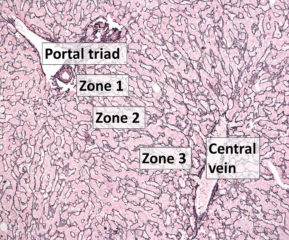
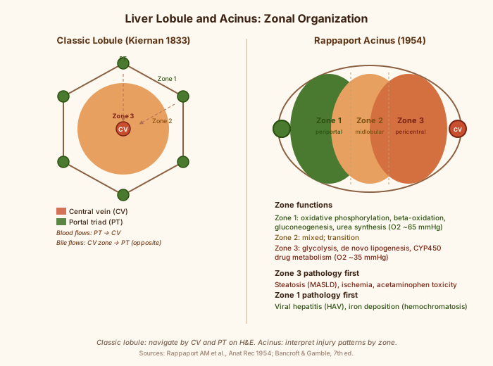
3. Lobule and Acinus: Two Models of the Same Organ

These are not competing theories. They describe different aspects of the same architecture. Knowing both is non-negotiable.

Model Classic lobule (Kiernan 1833) · Unit Hexagonal lobule · Center Central vein · Periphery Portal triads at corners · Best for Histologic orientation; what you see on H&E
Rappaport AM et al., Anat Rec 1954
Model Rappaport acinus (1954) · Unit Oval acinus around terminal portal venule · Center Portal triad (Zone 1) · Periphery Central vein region (Zone 3) · Best for Functional and pathologic zonation; injury patterns
Rappaport AM et al., Anat Rec 1954

On H&E you navigate by the classic lobule: find the central vein, find the portal triads. When you interpret injury (who got hurt first?), switch to the acinus model: Zone 1 is the oxygen-rich periportal end, Zone 3 is the oxygen-poor pericentral end.

Term Center · Lobule model Central vein · Acinus model Portal triad (Zone 1)
Rappaport AM et al., Anat Rec 1954
Term Periphery · Lobule model Portal triad · Acinus model Central vein region (Zone 3)
Rappaport AM et al., Anat Rec 1954
Term Steatosis first · Lobule model Centrilobular · Acinus model Zone 3 (acinus): same location, different vocabulary
Bancroft & Gamble, 7th ed.
Term Viral hepatitis necrosis (HAV) · Lobule model Periportal · Acinus model Zone 1 (acinus): same location
Bancroft & Gamble, 7th ed.

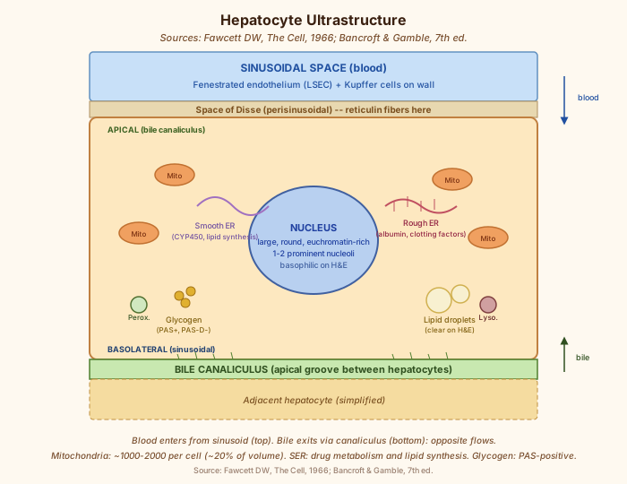
4. Ultrastructure

The hepatocyte is one of the most metabolically active cells in the body. Its ultrastructure reflects that.

Organelle / Feature Nucleus · Detail Large, round, central, 1-2 prominent nucleoli; euchromatin-rich (active transcription) · Histologic correlate Basophilic on H&E
Fawcett DW, The Cell, 1966
Organelle / Feature Smooth ER (SER) · Detail Abundant; site of CYP450 drug metabolism, lipid synthesis, glycogen storage · Histologic correlate PAS-positive cytoplasm; feathery change in cholestasis
Fawcett DW, The Cell, 1966
Organelle / Feature Rough ER (RER) · Detail Albumin, clotting factors, complement synthesis · Histologic correlate Basophilic cytoplasmic areas
Fawcett DW, The Cell, 1966
Organelle / Feature Mitochondria · Detail Most numerous organelle; 1000-2000 per cell in rat liver; ~20% of cell volume in rat · Histologic correlate Eosinophilic granular cytoplasm
Organelle / Feature Glycogen granules · Detail Alpha particles (rosettes) bound to smooth ER membranes through both deposition and depletion; centrilobular glycogen areas are infiltrated by SER tubules while periportal masses have SER only at the periphery (a structural difference, not an abundance gradient) · Histologic correlate PAS-positive; cleared by PAS-D
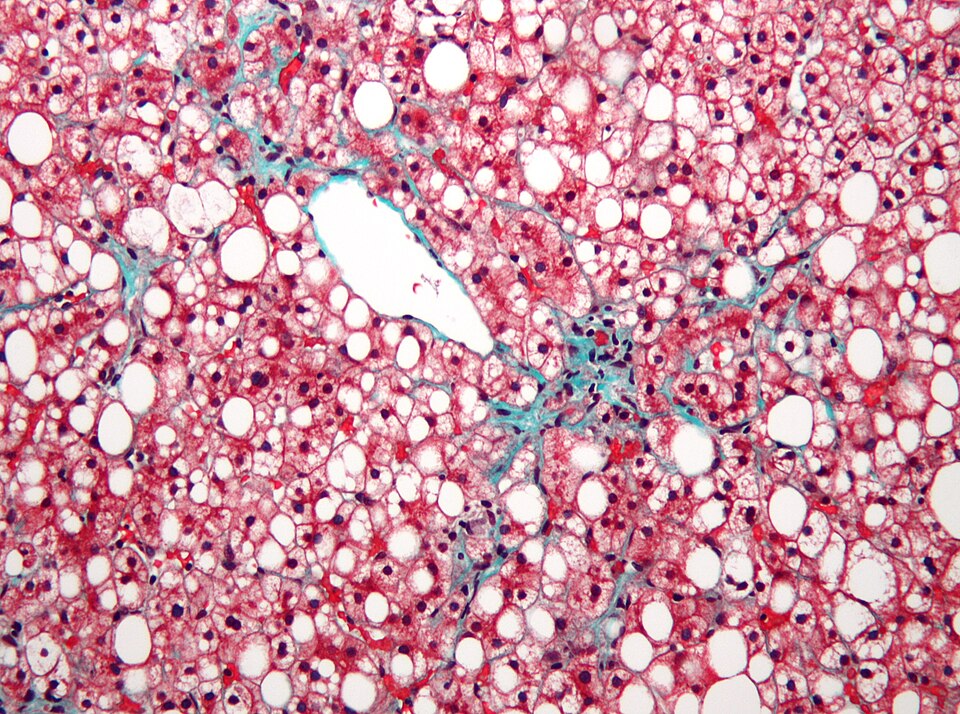
Organelle / Feature Lipid droplets · Detail Normally minimal; enlarged in steatosis · Histologic correlate Clear vacuoles on H&E; macrovesicular = one large, microvesicular = many small
Organelle / Feature Bile canaliculus · Detail Apical groove between adjacent hepatocytes; lined with microvilli; sealed by tight junctions · Histologic correlate Visible as clear space on H&E; dilated in cholestasis
Organelle / Feature Space of Disse · Detail Perisinusoidal space between hepatocyte basolateral surface and sinusoidal endothelium · Histologic correlate Reticulin fibers visible here; widened in perisinusoidal fibrosis (MASLD)
Organelle / Feature Peroxisomes · Detail Beta-oxidation of very-long-chain fatty acids; 300-600 per hepatocyte is quoted (thin: figure from a history review, no primary count cited) · Histologic correlate Not directly visible on routine stains
Organelle / Feature Lysosomes · Detail Lipofuscin accumulation (brown granules) with age · Histologic correlate Brown pigment on H&E; PAS-positive (diastase-resistant)

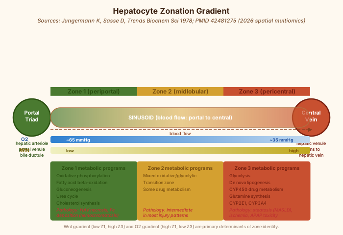
5. Zonation (Lobular Organization)

The hepatic lobule is a gradient organ. Every hepatocyte's gene program is tuned to its position along the portal-to-central axis.

Zone Zone 1 (periportal) · Location Around portal triads · Oxygen High (~65 mmHg) · Key metabolic programs Oxidative phosphorylation, beta-oxidation, gluconeogenesis, urea cycle, cholesterol synthesis · Pathology first affected Viral hepatitis necrosis (HAV, yellow fever); insulin resistance in MASLD; periportal iron in hereditary hemochromatosis
Jungermann K, Sasse D 1978
Zone Zone 2 (midlobular) · Location Intermediate · Oxygen ~45 mmHg · Key metabolic programs Mixed; transition zone · Pathology first affected Intermediate in most injury patterns
Jungermann K, Sasse D 1978
Zone Zone 3 (pericentral / centrilobular) · Location Around central veins · Oxygen Low (~35 mmHg) · Key metabolic programs Glycolysis, lipid synthesis (de novo lipogenesis), CYP450 drug metabolism, glutamine synthesis · Pathology first affected Steatosis (MASLD); ischemic/congestive necrosis; drug toxicity (acetaminophen); Zone 3-first bridging fibrosis

Molecular basis of zonation: Wnt signaling gradient (highest pericentral, lowest periportal) drives Zone 3 identity. Oxygen gradient (highest periportal) drives Zone 1 oxidative metabolism. Bile acid gradient provides additional positional information. All three are mapped in the 2026 spatial multiomics reference atlas (Ramachandran P, https://pubmed.ncbi.nlm.nih.gov/42481275/).

Regulators: Nr1i3 (nuclear receptor) and Nfix (transcription factor) control hepatocyte maturation and zone identity; CRISPR loss-of-function shifts zone gene programs (thin: single study, mouse; Stephan TL 2026, https://pubmed.ncbi.nlm.nih.gov/41819069/). Pericentral LSEC c-Kit activates FGF1 via RXRG, suppressing hepatocyte lipid accumulation through FGFR4; disrupting this paracrine axis is an early event in Zone 3 steatosis (thin: single study, mouse; Fang Z 2026, https://pubmed.ncbi.nlm.nih.gov/42319926/).

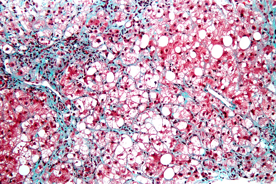
Zone 3 steatosis is the signature of early MASLD: pericentral hepatocytes are more lipogenic and more hypoxia-sensitive. A trichrome shows Zone 3 bridging fibrosis (centrilobular-to-centrilobular) before portal-to-portal bridging appears in many MASLD cases. This is the histologic readout of a molecular gradient being overwhelmed.

6. Pathology

Classic hepatocyte lesions and their histological appearance.

Lesion Steatosis, macrovesicular · Histologic appearance Single large clear vacuole displaces nucleus to periphery · Stain best for it H&E; Oil Red O (frozen only) · Zonation Zone 3 first (MASLD); Zone 1 first (some toxic)
Lesion Steatosis, microvesicular · Histologic appearance Multiple tiny vacuoles; nucleus central; more acute and ominous · Stain best for it H&E · Zonation Pan-lobular in severe cases
Lesion Hepatocyte ballooning · Histologic appearance Enlarged, pale, wispy cytoplasm; collapsed intermediate filaments; surrogate for cell stress; precedes Mallory-Denk bodies · Stain best for it H&E; reticulin shows plate collapse · Zonation Zone 3 in MASLD
Lesion Mallory-Denk bodies · Histologic appearance Eosinophilic cytoplasmic inclusions; ubiquitinated intermediate filament aggregates; in ballooned cells · Stain best for it H&E; CK8/18 IHC confirms · Zonation Zone 3
Standard hepatology reference
Lesion Hepatocyte necrosis (apoptosis/lytic) · Histologic appearance Acidophil bodies (apoptosis: pyknotic nucleus, eosinophilic cytoplasm); lytic necrosis: ghost cells or empty space · Stain best for it H&E · Zonation Zone 1: viral, ischemia in some; Zone 3: drugs, ischemia
Bancroft & Gamble, 7th ed.
Lesion Bridging necrosis · Histologic appearance Confluent necrosis connecting vascular structures; may be portal-central (most severe) · Stain best for it H&E; reticulin shows collapse bands · Zonation Zone 3 to Zone 1 in severe cases
Bancroft & Gamble, 7th ed.
Lesion Fibrosis (perisinusoidal) · Histologic appearance "Chicken wire" collagen in space of Disse; earliest MASLD fibrosis · Stain best for it Masson trichrome (blue); reticulin (condensed black) · Zonation Zone 3
Lesion Fibrosis (portal/bridging) · Histologic appearance Portal fibrosis, portal-portal bridges, portal-central bridges, cirrhosis · Stain best for it Masson trichrome · Zonation Progressive; cirrhosis = pan-lobular
Lesion Cirrhosis · Histologic appearance Diffuse fibrosis with regenerative nodules; lobular architecture lost · Stain best for it Trichrome; reticulin shows nodule borders without normal plate pattern · Zonation Pan-lobular
Bancroft & Gamble, 7th ed.
Lesion Glycogen accumulation · Histologic appearance Pale cytoplasm or clear on H&E; magenta on PAS; cleared on PAS-D · Stain best for it PAS; PAS-D · Zonation Zonal distribution: no abundance gradient is sourced. Lefkowitch 17355092 does not mention zone or glycogen; Babcock 1180230 reports a structural difference in SER arrangement, not differing amounts
Lesion Iron overload (hemosiderosis) · Histologic appearance Golden-brown granules in cytoplasm on H&E; blue-green on Perls · Stain best for it Perls Prussian blue · Zonation Zone 1 in hereditary hemochromatosis; diffuse in secondary overload
Lesion Copper accumulation · Histologic appearance Orange-brown granules on rhodanine; black on orcein; in Wilson disease and chronic cholestasis · Stain best for it Rhodanine; orcein · Zonation Periportal in Wilson; periportal in cholestasis
Lesion Alpha-1-antitrypsin globules · Histologic appearance Periportal PAS-positive, diastase-resistant magenta globules; globules persist after PAS-D · Stain best for it PAS-D (definitive) · Zonation Periportal (Zone 1)
AASLD reference
Lesion Ground-glass hepatocytes · Histologic appearance Finely granular pale cytoplasm; in HBV infection (HBsAg) or drug effect · Stain best for it Orcein (HBsAg); H&E for morphology · Zonation Periportal in HBV
Lesion Cholestasis · Histologic appearance Bile plugs in dilated canaliculi; feathery degeneration of periportal hepatocytes (bile salt toxicity) · Stain best for it H&E; Hall method (bile: green) · Zonation Zone 3 canalicular; Zone 1 hepatocellular in severe
Bancroft & Gamble, 7th ed.

7. Methods and Stains (as Knowledge)

The chemistry is what matters here.

Stain H&E · Principle Hematoxylin (basic dye) binds nucleic acids; eosin (acid dye) binds cytoplasmic proteins · Target in liver Nuclei blue, cytoplasm pink, RBCs red · Expected result Standard · Key interpretation Baseline morphology: steatosis, necrosis, inflammation, ballooning, cholestasis
Lab standard
Stain Masson trichrome · Principle Iron hematoxylin (nuclei); Biebrich scarlet/acid fuchsin (cytoplasm/muscle); phosphomolybdic-phosphotungstic acid displaces red from porous collagen; aniline blue (collagen) · Target in liver Collagen · Expected result Nuclei black, cytoplasm/muscle red, collagen blue · Key interpretation Fibrosis staging; perisinusoidal "chicken wire" = Zone 3 MASLD; bridging = advanced disease
Bancroft & Gamble, 7th ed.; https://pubmed.ncbi.nlm.nih.gov/17355092/
Stain Reticulin (Gordon-Sweet) · Principle Silver impregnation of type III collagen fibers · Target in liver Reticulin framework · Expected result Black fibers on pale background · Key interpretation Normal: one-cell-thick plates; HCC: plate thickening + fiber loss; collapse: condensed black bands; NRH: compressed borders
Stain PAS · Principle Periodic acid oxidizes vicinal diols in polysaccharides to aldehydes; Schiff reagent reacts with aldehydes to give magenta · Target in liver Glycogen, mucin, alpha-1-AT globules, basement membranes · Expected result Magenta-positive structures · Key interpretation Glycogen assessment; PAS-positive periportal globules suggest alpha-1-AT (confirm with PAS-D)
Stain PAS-D · Principle Diastase (amylase) pre-digests glycogen before PAS; diastase-resistant material remains magenta · Target in liver Alpha-1-AT globules, fungi, some basement membranes · Expected result Glycogen cleared; true deposits remain · Key interpretation Alpha-1-AT globules: magenta after PAS-D; glycogen: cleared; difference = the diagnosis
AASLD reference
Stain Perls iron (Prussian blue) · Principle Ferric iron reacts with potassium ferrocyanide at low pH to give blue-green precipitate (Berlin blue) · Target in liver Hemosiderin · Expected result Blue-green granules · Key interpretation Grading iron overload: 0-4 scale; periportal distribution in hereditary hemochromatosis; diffuse in transfusion overload
Stain Rhodanine · Principle Rhodanine chelates copper to form orange-red precipitate · Target in liver Copper · Expected result Orange-brown granules · Key interpretation Wilson disease; chronic cholestasis; periportal distribution; less sensitive than orcein for some deposits
Stain Orcein (Victoria blue) · Principle Orcein binds copper-associated protein (metallothionein-bound); also binds HBsAg in ground-glass hepatocytes · Target in liver Copper-associated protein; HBsAg · Expected result Brown-black granules · Key interpretation Wilson disease; chronic cholestatic conditions; HBsAg detection (complementary to IHC); periportal distribution
Stain Hall (bile stain) · Principle Fouchet reagent oxidizes bilirubin to biliverdin (green); counterstain highlights background · Target in liver Bile · Expected result Green bile plugs · Key interpretation Cholestasis confirmation; canalicular plugs in Zone 3; bile infarcts
Bancroft & Gamble, 7th ed.
Stain Oil Red O · Principle Dissolves in neutral lipid droplets; requires frozen section · Target in liver Lipid · Expected result Red-orange droplets · Key interpretation Confirms lipid; cannot be used on paraffin-processed tissue (lipid is extracted by solvents)
Bancroft & Gamble, 7th ed.

8. Regeneration

The liver is the only solid organ that can regenerate to its original mass after major resection. That capacity lives in the hepatocyte.

Aspect Capacity · Detail Regeneration to ~100% original mass after 70% partial hepatectomy (70% hepatectomy model in rodents; clinically relevant for living-donor transplant)
Bancroft & Gamble, 7th ed.
Aspect Hepatocyte entry into cell cycle · Detail Normally quiescent (G0); after injury, hepatocytes enter G1 within hours; peak DNA synthesis at 24-36 hours in rodent PHx model
Michalopoulos GK, DeFrances M, Science 1997
Aspect Growth factors · Detail HGF (hepatocyte growth factor, from stellate cells) and EGF are primary mitogens; TNF and IL-6 act as priming cytokines
Michalopoulos GK, DeFrances M, Science 1997
Aspect Polyploidy · Detail Adult hepatocytes are commonly binucleate or polyploid (4N, 8N, 16N); these cells re-enter mitosis and contribute daughter cells; polyploidy does not prevent regeneration
Aspect Mitotic fidelity · Detail NEURL1B (E3 ubiquitin ligase) stabilizes microtubule-organizing centers during hepatocyte mitosis; CRISPR loss-of-function impairs mitotic accuracy (thin: single study, mouse)
Aspect Metabolic-proliferative coupling · Detail Hepatocyte proliferation competes with lipid metabolism for cellular resources; EGFR signalling separates proliferative from lipogenic programs and accelerates regeneration after major hepatectomy (thin: single study)
Aspect Cytokine orchestration · Detail Regeneration is not a simple proliferative response; requires inflammatory priming (TNF, IL-6), complement activation, mechanosensory signaling (portal pressure changes), and metabolic reprogramming
Aspect Architectural restoration · Detail PAR3-mediated polarity coordinates proliferation with lobular architecture restoration; ANXA2+ migratory hepatocytes close wounds and re-establish zonation (thin: single study)
Aspect Reprogramming as therapy · Detail Partial OSK (Oct4/Sox2/Klf4) mRNA reprogramming via lipid nanoparticles to hepatocytes reverses fibrosis markers in mouse models; thin: single study, mouse; clinical translation pending
Aspect In vitro barrier · Detail Primary hepatocytes lose differentiated function rapidly in standard culture; no reliable clinical-grade expansion protocol yet; 3D organoid systems and co-culture with nonparenchymal cells are the current frontier

Histologic correlate: during active regeneration, reticulin stain shows restoration of one-cell-thick plate architecture as new trabeculae form. Trichrome shows collagen regression if the injury resolves. A regenerating liver that fails to restore reticulin architecture is not healing normally.

9. Reticulin Stain in Liver Pathology

Feature Fiber composition · Detail Reticulin = type III collagen; forms the hepatic trabecular scaffold (one-cell-thick plates), sinusoidal lining, portal tract framework
Feature Normal architecture · Detail One-cell-thick hepatocyte plates; reticulin wraps each plate; sinusoids run between
Bancroft & Gamble, 7th ed.
Feature HCC pattern · Detail Plates become two or more cells thick (trabecular pattern); reticulin fibers lost or disrupted around nodules
Feature Pitfall: fatty liver · Detail Macrovesicular steatosis can cause reticulin loss in benign liver, mimicking HCC; never diagnose HCC on reticulin alone
Feature Pitfall: reticulin-intact HCC · Detail Rare well-differentiated HCCs retain reticulin; use glypican-3, Ki-67, and glutamine synthetase to resolve
Feature Collapse pattern · Detail Confluent reticulin condensation without hepatocytes = zonal necrosis with collapse; seen in ischemia, drug injury, acute hepatitis
Feature NRH · Detail Nodular regenerative hyperplasia: one-cell-thick plates expand nodularly; reticulin compressed at nodule borders; not HCC
PathologyOutlines (NRH)
Feature Triple stain · Detail Reticulin + glypican-3 + glutamine synthetase on one section differentiates HCC from hepatic adenoma from FNH
Feature MASLD/HCC context · Detail AI-assisted analysis of reticulin and H&E shows sex-specific patterns in MASLD-driven HCC

10. Hepatocyte Ballooning and Histologic Scoring

Ballooning is not steatosis. A ballooned hepatocyte is dying; a steatotic hepatocyte is storing fat. Getting this right on H&E is what separates a MASLD call from a MASH call.

Feature Vacuole · Ballooning Absent or diffuse cytoplasmic rarefaction · Macrovesicular steatosis Single large clear vacuole, well-defined
Feature Nucleus · Ballooning Central, may look displaced by swelling · Macrovesicular steatosis Pushed to cell periphery
Feature Cytoskeleton · Ballooning Intermediate filament collapse; MDB precursor · Macrovesicular steatosis Intact
Feature Reticulin · Ballooning Condensed around collapsed cell · Macrovesicular steatosis Normal plate architecture preserved
Feature Significance · Ballooning Necroinflammatory; MASH criterion · Macrovesicular steatosis Steatosis; may be MASLD without MASH

Scoring systems:

System NAS (NASH CRN; Kleiner 2005) · Components Steatosis 0-3, lobular inflammation 0-2, ballooning 0-2; total 0-7 · Notes NAS is an activity score, not a MASH diagnosis
System SAF (Bedossa 2012) · Components Steatosis 0-3, Activity (ballooning + lobular inflammation) 0-4, Fibrosis 0-4 · Notes Used in European trials; thin: which is superior is debated
Bedossa P, J Hepatol 2012
System Portal inflammation · Components Contested component; NASH CRN excludes it from grade; 2026 head-to-head still unresolved · Notes n/a
System Extended ballooning score · Components Subcategories added to 0-2 scale; improved interobserver agreement (thin: single study) · Notes n/a
System Digital/AI scoring · Components Reduces interobserver variability for steatosis, inflammation, and ballooning; validated against NAS (thin: single study, pending multi-center replication) · Notes n/a

Reversibility: paired biopsies after bariatric surgery show significant reduction in steatosis grade, NAS, and ballooning (thin: single study; Li M 2025, https://pubmed.ncbi.nlm.nih.gov/40844300/). The stain findings are not permanent if the injury resolves.

Mechanical basis: lipid droplets deform the nucleus and disrupt actin cytoskeletal networks before histologic ballooning is visible (thin: single study; https://www.ncbi.nlm.nih.gov/pmc/articles/PMC10120019/). The functional impairment precedes the stain finding.

11. Hepatocellular Carcinoma (HCC): Hepatocyte Perspective

Feature Cell of origin · Detail Hepatocyte or hepatocyte progenitor (debate ongoing; thin)
Standard hepatology
Feature Histologic diagnosis · Detail Trabecular, acinar, solid, or scirrhous patterns on H&E; reticulin loss is the key architectural clue
Feature Reticulin · Detail Lost or disrupted in most HCC; retained in rare well-differentiated tumors
Feature Immunohistochemistry panel · Detail HepPar1 (hepatocyte paraffin antigen), arginase-1, glypican-3 (positive in HCC), glutamine synthetase (diffuse in HCC)
Feature Triple stain · Detail Reticulin + glypican-3 + glutamine synthetase on one section covers most diagnostic scenarios
Feature MASLD-driven HCC · Detail Steatotic liver creates pro-metastatic niche (thin: single study); AI histology reveals sex-specific progression patterns (thin: single study)

12. Nonparenchymal Cells in Context

The hepatocyte does not work alone. These cells shape its function and its histological appearance.

Cell Hepatic stellate cell (Ito cell) · Location Space of Disse · Role Quiescent: vitamin A storage; activated: myofibroblast, collagen production (fibrosis) · Stain visibility Not visible on H&E in quiescent state; alpha-SMA IHC for activated; trichrome shows their product (collagen)
Cell Kupffer cell · Location Sinusoidal lumen · Role Resident macrophage; phagocytosis; inflammatory signaling · Stain visibility Plump cells on sinusoidal lining on H&E; CD68 IHC
Cell Liver sinusoidal endothelial cell (LSEC) · Location Sinusoidal wall · Role Fenestrated endothelium; zone-specific paracrine signaling (FGF1/FGFR4 axis in steatosis; thin: single study, mouse) · Stain visibility Lining cells on H&E; not distinguished from Kupffer without IHC
Cell Cholangiocyte · Location Bile ducts · Role Bile modification; bile duct reaction in injury; progenitor niche · Stain visibility Duct structures on H&E; CK7/CK19 IHC
Bancroft & Gamble, 7th ed.

MASLD reframe: nonparenchymal cells direct hepatocyte function. The fibrosis seen on trichrome reflects HSC activation, not a hepatocyte-intrinsic problem. LSEC zonation signals set the Zone 3 lipogenic program. Hepatocyte pathology must be read in its stromal context (thin: single study; Jaeger JW 2026, https://pubmed.ncbi.nlm.nih.gov/42342151/).

13. Recent Findings (2026)

Finding High-resolution spatial multiomics: human liver zonation reference atlas · Significance Wnt, oxygen, and bile acid gradients mapped at single-cell resolution; the molecular recipe behind every histologic pattern
Finding LSEC c-Kit / FGF1 / FGFR4 axis drives Zone 3 steatosis onset · Significance Endothelial-hepatocyte paracrine signaling sets pericentral lipogenic identity; not hepatocyte-intrinsic alone (thin: single study, mouse)
Finding Nr1i3 and Nfix regulate hepatocyte maturation and zonation · Significance CRISPR screen; loss shifts zone identity (thin: single study, mouse)
Finding PAR3 / ANXA2 restores lobular architecture during regeneration · Significance Polarity protein coordinates migratory hepatocyte wound closure and zone re-establishment (thin: single study)
Finding NEURL1B regulates mitotic fidelity in regenerating hepatocytes · Significance E3 ligase stabilizes spindle assembly; CRISPR loss impairs mitosis (thin: single study, mouse)
Finding OSK mRNA reprogramming reverses hepatocyte fibrosis · Significance Partial cellular reprogramming via LNPs; thin: single study, mouse; clinical translation pending
Finding Updated MASLD histologic criteria · Significance Refined definitions for ballooning, lobular inflammation, portal changes; affects scoring
Finding Nonparenchymal cells orchestrate hepatocyte metabolism in steatotic liver · Significance Stromal context is inseparable from hepatocyte pathology (thin: single study)
Finding Digital/AI scoring reduces interobserver variability · Significance NAS and ballooning assessment improved by machine learning (thin: single study)
Finding MASLD creates pro-metastatic hepatic niche · Significance Altered zonation, oxygen gradients, immune changes in steatotic liver favor tumor seeding (thin: single study)
Finding Bariatric surgery reverses MASLD histology on paired biopsies · Significance Steatosis grade, NAS, and ballooning significantly reduced post-surgery (thin: single study)
Finding STING is an organelle-stress checkpoint in hepatocytes · Significance Sensing mitochondrial and ER damage, STING drives hepatocyte fate: survival, pyroptosis, or fibrogenic remodeling; extends STING biology beyond antiviral defense (thin: single study)
Finding Hepatocyte-HSC bidirectional paracrine crosstalk drives fibrosis · Significance Hepatocyte-derived TGF-beta and lipid mediators activate HSCs; activated HSCs return pro-fibrotic and pro-apoptotic signals; fibrosis is a two-way conversation (thin: single study)
Finding Ubiquitin-immune axis links metabolic stress to hepatocyte fate across liver diseases · Significance Ubiquitin-proteasome dysregulation in hepatocytes converges MASLD, viral hepatitis, and HCC pathogenesis; proposed as a unifying therapeutic target (thin: single review, pending primary replication)
Finding hESC-derived polarized hepatocyte organoids recapitulate MASLD and enable drug screening · Significance Mature, polarized organoids develop steatosis and lipotoxicity in FFA medium; validated for compound screening (thin: single study)
Finding Extracellular vesicle signatures distinguish PHH, organoid, and patient serum in MASH · Significance Each model captures distinct EV biology; complementary use required for biomarker discovery (thin: single study)
Finding Oxygenated perfusion rescues CYP450 and albumin in iPSC-liver tissue · Significance Controlled O2 in bioreactor addresses Zone 3 hypoxia in static culture; key for drug metabolism assays (thin: single study)
Finding Spatial immune dysregulation in MASLD integrates lobular zoning with immune function · Significance Spatial transcriptomics maps zone-specific immune cell composition and pro-inflammatory signaling in MASLD; perivenular (Zone 3) injury drives the earliest immune activation pattern (thin: single study)
Finding Immune metabolic reprogramming in MASLD: Kupffer cell and lymphocyte reprogramming by steatosis · Significance Lipotoxicity, ER stress, and altered bile acids reprogram hepatic immune cells; links zone-3 lipid accumulation to perivenular inflammation and disease progression (thin: review)
Latest 8

2026-09-09 · PubMed
Redefining healthy human liver zonation using high-resolution spatial multiomics

High-resolution spatial transcriptomics and proteomics map the full zonation gradient in healthy human liver; establishes reference atlas for normal at the molecular level before disease.

2026-09-09 · PubMed
PAR3-mediated coordination of hepatocyte proliferation, maturation, and architecture in regeneration

PAR3 polarity protein links hepatocyte proliferation to architectural restoration during regeneration; migratory ANXA2+ hepatocytes restore lobular zonation via wound-closure-mediated reorganization.

2026-09-09 · PubMed
CRISPR screen: E3 ligase NEURL1B regulates hepatocyte mitosis via microtubule-organizing centers

In vivo CRISPR interference screen in regenerating mouse liver identified NEURL1B as a regulator of mitotic spindle fidelity during hepatocyte division.

2026-09-09 · PubMed
AI-assisted histopathology for sex-specific MASLD-related HCC

AI-enhanced image analysis of H&E and special stains reveals sex-specific differences in MASLD-driven HCC progression and regression.

2026-09-09 · PubMed
MASLD reprograms the hepatic metastatic niche

Steatotic liver creates a pro-metastatic niche via altered zonation, oxygen gradients, lipid metabolism, and immune activity.

2026-09-09 · PubMed
Digital pathology tailored for liver biopsy assessment

AI-assisted scoring of liver biopsies for steatosis, inflammation, ballooning, and fibrosis; reduces interobserver variability on special stains.

2026-09-09 · PubMed
Triple stain reticulin/glypican-3/glutamine synthetase for liver lesions

Triple stain panel differentiates HCC, hepatic adenoma, and FNH on biopsy. Reticulin is the architectural anchor; GPC3 and GS add immunohistochemical specificity.

2026-09-09 · PubMed
Special stains in diagnostic liver pathology (Lefkowitch 2006)

Foundational review of trichrome, reticulin, PAS, iron, and copper stains in liver pathology. The primary bench reference.