Gallery
hepatocyte images: illustrations and micrographs. All credits and licenses listed.
Micrographs
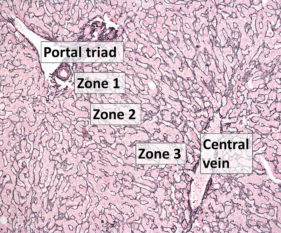
Normal human liver: reticulin stain with zones labelled. Portal triad (Zone 1) to central vein (Zone 3). Hepatocyte plates outlined in reticulin fibres.
Boyd A, Cain O, Chauhan A, Webb GJ · CC BY 4.0 · source
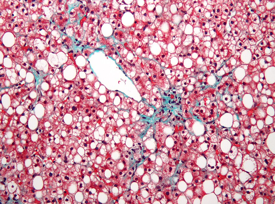
Macrovesicular steatosis in NAFLD/MASLD. Trichrome stain: large lipid vacuoles (white) displace hepatocyte nuclei; pericellular fibrosis in blue-green; central vein centre-right.
Nephron · CC BY-SA 3.0 · source
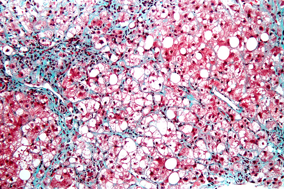
Steatohepatitis (MASH). Trichrome stain at high magnification: ballooned hepatocytes, microvesicular and macrovesicular steatosis, lobular inflammation, chicken-wire pericellular fibrosis.
Nephron · CC BY-SA 3.0 · source
Illustrations
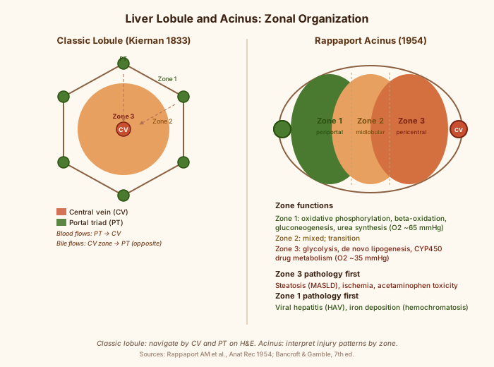
Classic lobule (Kiernan 1833) and Rappaport acinus (1954): zonal organization, vascular landmarks, and injury patterns by zone.
Illustration by hepato.site · CC0
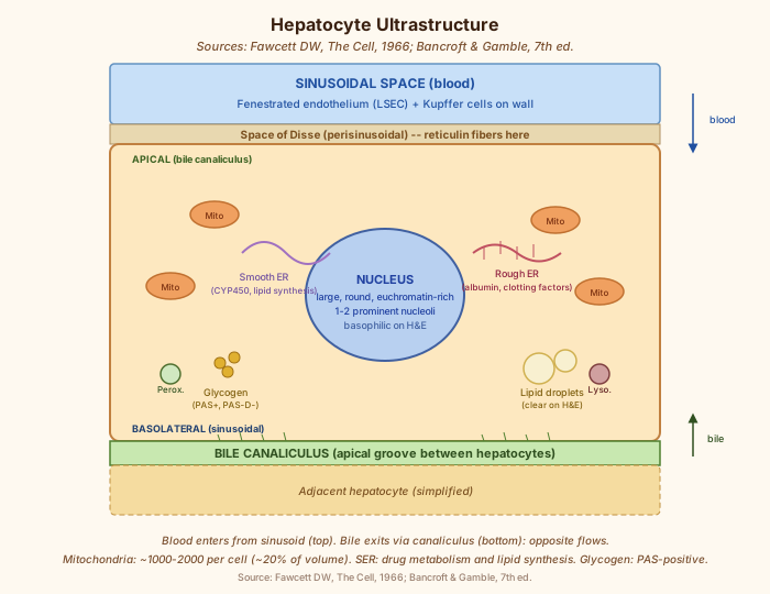
Hepatocyte ultrastructure: nucleus, mitochondria, smooth and rough ER, glycogen, lipid droplets, bile canaliculus, space of Disse. Sources: Fawcett 1966; Bancroft & Gamble 7th ed.
Illustration by hepato.site · CC0
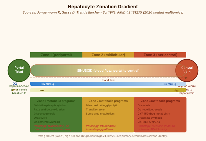
Hepatocyte zonation gradient from portal triad (Zone 1) to central vein (Zone 3): O2 gradient, Wnt gradient, metabolic programs by zone, and zone-specific pathology.
Illustration hepato.site; data after Jungermann K 1982 and Ramachandran P et al. 2026 · CC0